Savonia Article: VR for Anatomy Learning in Radiography Studies
This work is licensed under CC BY-SA 4.0


Virtual reality is opening new possibilities for radiography education by making complex anatomical structures more visible, interactive, and easier to understand.
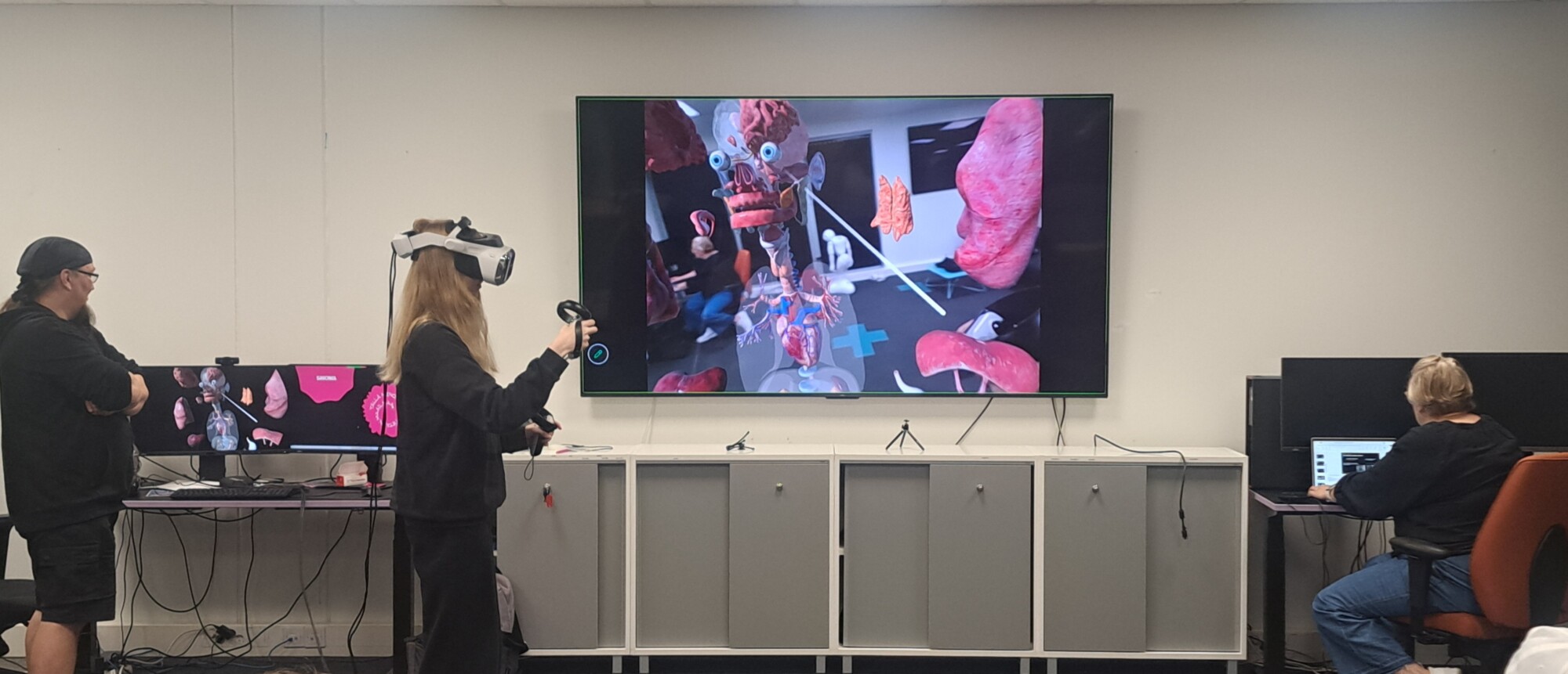
For the first time, second-year radiography students were introduced to the use of VR in anatomy learning as part of the course Computed Tomography Examinations and Interventions, 5 ECTS. The VR session complemented the practical workshops covered in the course. The session took place at the XR Center facilities and used a Varjo Medical Human Anatomy VR application (Varjo technical demo). Four students tested the VR equipment directly, while the rest of the group followed the teaching session online from different campuses through a satellite learning model. This made it possible to combine hands-on experimentation with remote participation.
The main aim of the VR session was to help students develop a stronger three-dimensional understanding of human anatomy and connect this knowledge directly with radiography practice. By exploring anatomical structures in an immersive environment, students could better understand how body structures are located in relation to each other and how this spatial knowledge supports imaging decisions, such as patient positioning, image centering, and identifying relevant body areas in computed tomography.
During the session, the teacher guided students through exercises involving anatomical structures relevant to radiography and imaging, including the hippocampus, tracheal bifurcation, aorta, pancreas, lumbar spine, nerve roots, and three-dimensional anatomical views. These examples helped students link anatomical knowledge to clinical imaging contexts and understand the role of anatomical landmarks in ensuring that all necessary body structures are included in diagnostic images.
Students’ first reactions to the VR session were mainly positive. They described the experience as interesting, engaging, and useful because it allowed them to move anatomical structures, view the body from different perspectives, and examine details that can be difficult to understand through textbooks or two-dimensional images alone. Some students also noted that using VR for the first time felt slightly intimidating, underlining the importance of clear instructions, calm guidance, and enough time to become familiar with the equipment.
The experience showed that VR can be a valuable complementary tool in radiography education. When used with clear learning objectives and guided reflection, VR can support anatomy learning, improve spatial understanding, and help students connect theoretical knowledge with future professional practice. In the future, VR-based anatomy materials could also be included in students’ learning portfolios, encouraging them to collect, compare, and reflect on different resources that support their development as radiography professionals.
For more information on how to incorporate VR into courses, please contact:
Anne-Mari Kulhomäki, Teacher Radiography, 044 785628, anne-mari.kulhomaki@savonia.fi
Kimmo Pakarinen, XR expert, 0447856034, Kimmo.pakarinen@savonia, XR center, https://xrcenter.savonia.fi/
Authors
Paola Kontro, RDI expert, Savonia University of Applied Sciences
This article was prepared with the help of AI.